



 0752-2228195
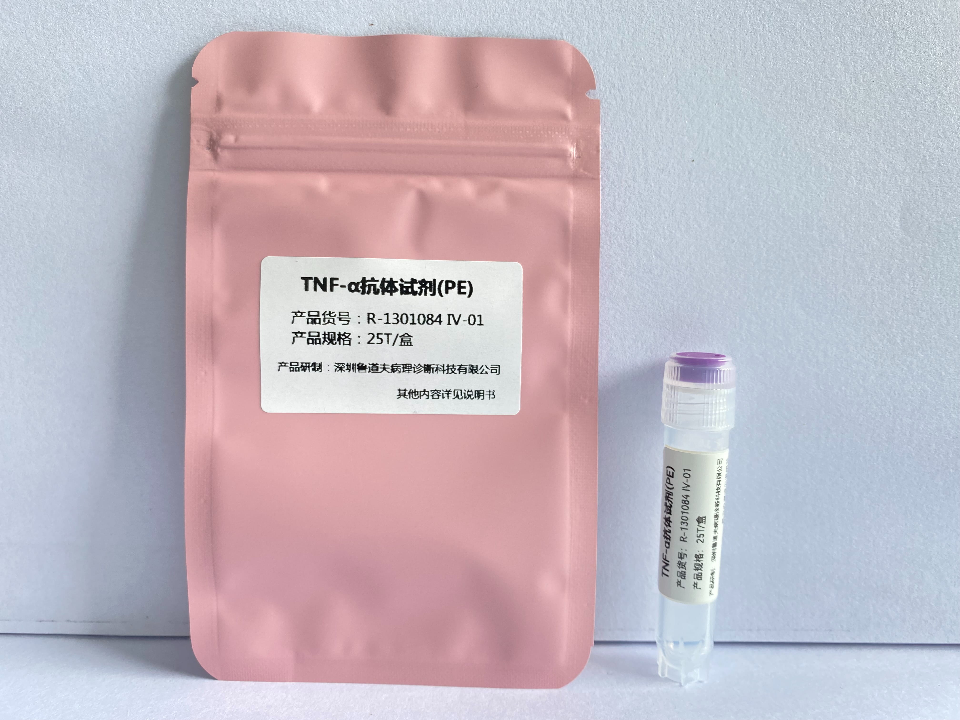
TNF-α antibody reagent (PE)
Product Code:R-1301084Ⅳ-01/R-1301084Ⅳ-02
Applicable Scenarios:For scientific use only.

| Product Name | Anti-Human TNF-α, PE Detection Reagent |
|---|---|
| Target | TNF, TNFA, TNFSF2 |
| Marker | PE |
| Instrument | Flow cytometer compatible with 488nm, 532nm, 561nm lasers |
| Subtype | Mouse IgG1, κ |
| Clone Number | MAb11 |
| Recommended Application | Flow cytometry |
| Dosage | 5 μL /Test |
| Purification | Affinity chromatography |
| Stock Solution | Phosphate buffer solution containing 0.2% BSA and 0.2% proclin 950 (pH7.2) |
| Storage Conditions | Store at 2 - 8℃, protect from light, do not freeze |
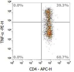
Fig.-After human peripheral blood was blocked by PMA/Ionomycin Mixture and BFA/Monensin Mixture for 4 hours, the membrane was fixed with a fixed membrane breaker, and Anti-Human TNF-α,PE (Clone: MAb11), Anti-Human CD4, APC APC were stained. The CD3+ cells in FSC/SSC were gated for analysis.
Overview of TNF molecular target information
Molecular name: TNF, tumor necrosis factor
Gene family: Tumor necrosis factor superfamily
Alias: TNFSF2; DIF; TNF-alpha
Former name: TNFA
Full name: TNF superfamily, member 2; tumor necrosis factor (TNF superfamily, member 2)
Review of TNF molecular targets
TNF-α is a 17 kDa cytokine produced by monocytes, macrophages, neutrophils, NK cells and CD4+T cells. It has many regulatory functions in immune response and can also be used as a potential pyrogen. After TNF-α responds to stimuli (infectious factors or tissue damage), it activates neutrophils, changes the characteristics of vascular endothelial cells, regulates the metabolic activity of other tissues, and demonstrates tumor killing activity by inducing local coagulation. TNF-α plays an important role in the pathogenesis of inflammation in joint tissues and other tissues.




Copyright: Shenzhen Rudolf Pathological Diagnosis Technology Co., Ltd